2023 FDA Science Forum
Assessing the Developmental Effects of Fentanyl (Anesthetics/Analgesics) Using Neural Stem Cell Models
- Authors:
- Center:
-
Contributing OfficeNational Center for Toxicological Research
Abstract
Fentanyl is a potent and short-acting opioid medication that is often given to pediatric patients during surgery to relieve pain and as an adjunct to an anesthesia. While it is difficult to verify the adverse anesthetic effects on human infants and children, the utilization of highly relevant preclinical models, such as neural stem cells derived from humans, might serve as a “bridging” model to evaluate the vulnerability of the developing nervous system and address FDA’s regulatory needs.
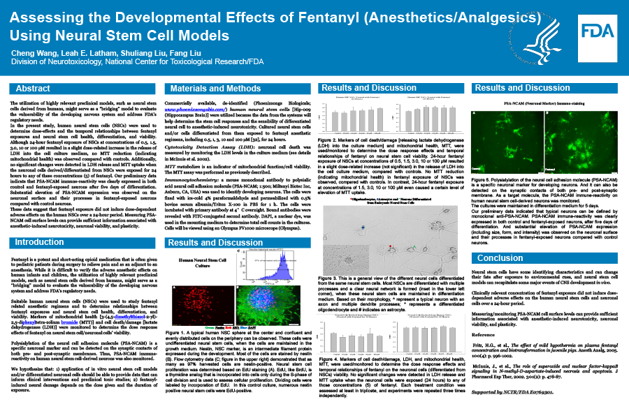
In the present study, human neural stem cells (NSCs) were used to determine dose-effects and the temporal relationships between fentanyl exposures and neural stem cell health, differentiation, and viability. Markers of mitochondrial health [3-(4,5-dimethylthiazol-2-yl)-2,5-diphenyltetra-zolium bromide (MTT)] and cell death/damage [lactate dehydrogenase (LDH)] were monitored to determine the dose response effects of fentanyl on neural stem cell/neuronal cells’ viability. Although 24-hour fentanyl exposure of NSCs at concentrations of 0.5, 1.5, 3.0, 10 or 100 µM resulted in a dose-related increase (not significant) in the release of LDH into the cell culture medium, no significant MTT reduction (indicating mitochondrial health) was observed compared with controls. Additionally, no significant changes were detected in MTT uptake when the neuronal cells derived/differentiated from NSCs were exposed for 24 hours to any of these concentrations (5) of fentanyl. Polysialylation of the neural cell adhesion molecule (PSA-NCAM) immune-reactivity on human neural stem cell-derived neurons was also monitored. PSA-NCAM is a specific neuronal marker and can be detected on the synaptic contacts of both pre- and post-synaptic membranes. Our preliminary data indicate that PSA-NCAM immune-reactivity was clearly expressed in both control and fentanyl-exposed neurons after five days of differentiation. Substantial elevation of PSA-NCAM expression was observed on the neuronal surface and their processes in fentanyl-exposed neurons compared with control neurons.
These data suggest that clinically relevant concentrations of fentanyl exposure did not induce dose-dependent adverse effects on the human NSCs and neuronal cells over a 24-hour period. Measuring PSA-NCAM cell surface levels can provide sufficient information associated with anesthetic-induced neurotoxicity, neuronal viability, and plasticity.

