2023 FDA Science Forum
The development of an in vitro qPCR assay to detect and quantify HCoV-NL-63
- Authors:
- Center:
-
Contributing OfficeNational Center for Toxicological Research
Abstract
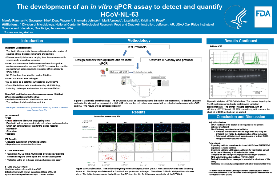
The family Coronaviridae houses etiological agents capable of causing clinical diseases in humans and animals, with the severity in humans ranging from the common cold to severe acute respiratory syndrome. Like SARS-CoV2, NL-63 invades host cells through the angiotensin converting enzyme-2 (ACE2), and the resulting mechanism of action may result in cytopathic effects similar to SARS-CoV2. However, the respiratory disease caused by NL-63 is milder, less infective, and self-limiting. As a result, NL-63 is a BSL-2 level pathogen, yet it could be a potential surrogate for SARS-CoV2. Current limitations exist in understanding NL-63 biology, including challenges in virus detection and quantitation. Many classical techniques, such as the plaque forming assays, are not reliable due to the uncharacteristic cytopathic effects of NL-63. To enhance the ability to quantitate NL-63, we developed a multiplexed qPCR assay targeting conserved regions of the spike and nucleocapsid genes, which we then validated using an in-house immunofluorescence assay. Virus was propagated in LLC-MK2 cells, quantified, and stored at -80 °C until the assay was performed. Briefly, 24 hours prior to inoculation, 1 x 10⁴ LLC-MK2 cells were seeded per well into a 96-well flat-bottomed plate treated with poly-lysine. The cells were allowed to adhere at 37 °C, confluency above 90% was confirmed, and 1 x 10⁵ FFU were inoculated into each well except for the negative control wells. Supernatants were collected 24 hours post-inoculation and the immunofluorescence assay was performed on the remaining cells with DAPI as a nucleus stain and GFP conjugated to an antibody targeting the NL-63 spike protein. Regarding the qPCR, the amplification efficiency of the qPCR assay was determined and consistently performed at 90 to 110%, with melt curves indicating a lack of non-specific amplifications and dimer formation. Preliminary data indicates that while detection appears successful across both assays, quantitating the virus at 1x10⁴ FFU, there are variable differences between the assays’ ability to quantitate the virus. However, both assays may correlate but it is evident they measure two very different metrics and both kinds of data taken together overcome limitations with earlier attempts at viral quantification.

