2023 FDA Science Forum
MALDI imaging mass spectrometry of mouse fetuses to assess markers of neural tube defects after maternal opioid exposure
- Authors:
- Center:
-
Contributing OfficeNational Center for Toxicological Research
Abstract
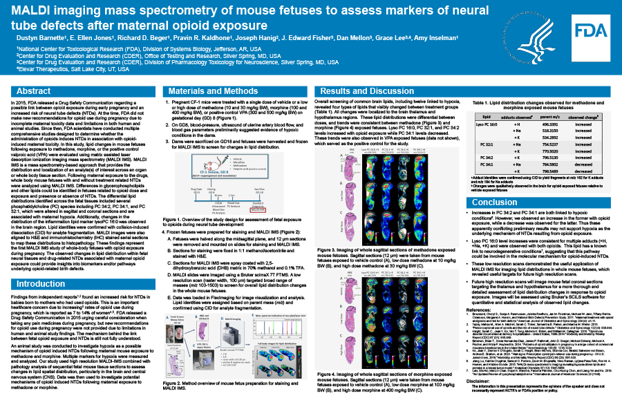
In 2015, FDA released a Drug Safety Communication regarding the possible link between opioid exposure during early pregnancy and an increased risk of neural tube defects (NTDs). At the time, FDA did not make new recommendations for opioid use during pregnancy due to limitations in both human and animal studies and incomplete maternal toxicity data. Since then, FDA scientists have conducted multiple comprehensive studies designed to determine whether the administration of opioids induces NTDs in association with maternal toxicity related to opioid exposure. In this study, fetal effects in mice following exposure to methadone, morphine, and the positive control valproic acid were evaluated using matrix assisted laser desorption ionization imaging mass spectrometry (MALDI IMS). MALDI IMS is a mass spectrometry-based approach that provides the distribution and localization of an analyte(s) of interest across an organ or whole body a tissue section. Thus, following maternal exposure to opioids, whole body mouse fetuses with and without treatment related NTDs were analyzed using MALDI IMS. Many differences in glycerophospholipids and other interesting lipids could be identified in fetuses related to opioid dose and exposure and presence or absence of NTDs. The differential lipid distributions identified across the fetal tissues included several phosphatidylcholine species such as PC 34:2, PC 36:1, and PC 34:1, which were altered in sagittal and coronal sections and are associated with maternal hypoxia. Additionally changes in the distribution of the inflammation lipid marker LysoPC 16:0 was also observed in the brain region. Lipid identities were confirmed with collision-induced dissociation for analyte fragmentation. MALDI images were also aligned to H&E and IHC stained serial sections to map these distributions to histopathology. These findings represent the first MALDI IMS study of whole-body fetuses with opioid exposure during pregnancy. The observed changes in lipid distribution within fetal neural tissues and drug-related NTDs associated with maternal opioid exposure could provide insights into biomarkers and/or pathways underlying opioid-related birth defects.

