2023 FDA Science Forum
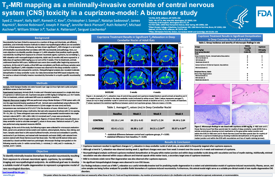
T2-MRI mapping as a minimally-invasive correlate of central nervous system (CNS) toxicity in a cuprizone-model: A biomarker study.
- Authors:
- Center:
-
Contributing OfficeNational Center for Toxicological Research
Abstract
Neurotoxicity has been linked to exposure to a number of drugs and chemicals, yet efficient, predictive, and minimally-invasive methods to detect neuroanatomical effects are not routinely used currently in non-clinical assessments. Previously, we have shown significant T2-MRI changes in a rat model of trimethyltin neurotoxicity that correlated with CNS neurotoxicity and pathology as measured by Flouro Jade C. Here, our main objective was to identify possible T2-MRI relaxation that predicts myelin specific neurotoxicity resulting from exposure to a known neurotoxic agent, cuprizone, by correlating T2-MRI relaxation with neuropathological endpoints. Adult-male rats (3-months old) were exposed to a daily dose of cuprizone (600 mg/kg p.o., daily) or corn oil for 4 weeks. Prior to treatment, animals underwent MRI scans to establish a baseline. Additional MRI scans were done weekly after beginning exposure to cuprizone. At the end of 4 weeks, a final MRI was completed and fluid and tissue samples were collected. Significant T2-MRI relaxation was observed in the deep cerebellar nuclei in cuprizone-treated rats compared to controls. A correlation of these MRI changes with CNS pathology will be discussed. Our data demonstrate that MRI-based endpoints may be used as a robust minimally-invasive neurotoxicity biomarker in a myelin-specific neurotoxicity model. Disclaimer: This presentation reflects the views of the authors and does not necessarily reflect those of the Food and Drug Administration.

